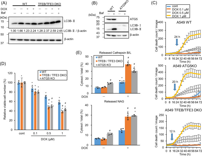
Fig. 3. TFEB/TFE3 induces autophagy and suppresses lysosomal membrane permeabilization (LMP) to protect cells from doxorubicin (DOX)-induced cell death.
A LC3B expression in wild-type (WT) or TFEB/TFE3 double knockout (DKO) A549 cells treated with 1 µM DOX for 24 h was assessed by western blotting in the presence or absence of 50 nM Bafilomycin A1 (Baf) for 2 h. β-actin was used as a control. The relative LC3B-II band intensity is shown. B ATG5-knockout (KO) was confirmed by western blotting. WT and ATG5-KO A549 cells were treated with/without 50 nM Baf for 24 h. β-actin was used as the loading control. C WT, ATG5-KO, and TFEB/TFE3 DKO A549 cells were treated with DOX and the dead cell count was monitored every 4 h by live-cell imaging system with propidium iodide (PI) staining. The cell death initiation point after 1 µM DOX treatment is indicated by blue arrows. D The viable WT, ATG5-KO, and TFEB/TFE3 DKO A549 cell count 24 h after DOX treatment. n = 4, bar = mean ± SD, *p < 0.05 vs. cont., #p < 0.05 vs. WT. E LMP in WT, ATG5-KO, and TFEB/TFE3 DKO A549 cells treated with 1 µM DOX for 24 h were measured by assessing released cytosolic N-acetyl glucosaminidase (NAG) or cathepsin B/L activity. n = 4, bar = mean ± SD, *p < 0.05 vs. cont., #p < 0.05 vs. WT.

