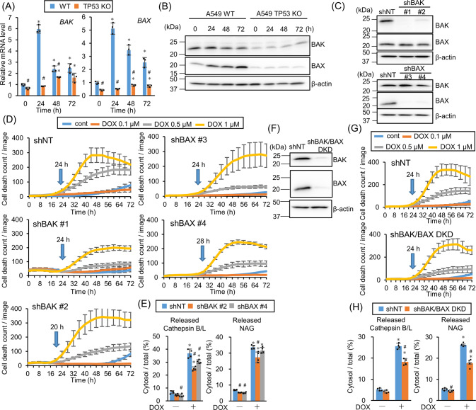
Fig. 4. BAK and BAX are partly involved in doxorubicin (DOX)-induced lysosomal membrane permeabilization (LMP).
A The relative BAK and BAX mRNA expression level in wild-type (WT) or TP53-knockout (KO) A549 cells treated with 1 µM DOX for 24, 48, or 72 h was assessed by real-time PCR. n = 3, bar = mean ± SD, *p < 0.05 vs. 0 h, #p < 0.05 vs. WT. B BAK and BAX expression in WT or TP53-KO A549 cells treated with 1 µM DOX for 24, 48, or 72 h were assessed by western blotting. β-actin was used as a loading control. C BAX and BAK knockdown (KD) efficiency in shRNA introduced A549 cells expressing shBAK or shBAX were confirmed by western blotting. β-actin was used as a control. D shNT-, shBAK #1-, #2-, or shBAX #3-, #4-expressing A549 cells were treated with DOX and the dead cell count was monitored every 4 h by live-cell imaging system with propidium iodide (PI) staining. E LMP in BAK- or BAX-KD A549 cells treated with 1 µM DOX for 24 h were measured by assessing released cytosolic N-acetyl glucosaminidase (NAG) or cathepsin B/L activity. n = 4, bar = mean ± SD, *p < 0.05 vs. cont., #p < 0.05 vs. shNT. F BAK and BAX KD efficiency in BAK/BAX double knockdown (DKD) cells was confirmed by western blotting. β-actin was used as a control. G Control or BAK/BAX DKD A549 cells were treated with DOX and dead cell count was monitored every 4 h by live-cell imaging system with PI staining. H LMP in shNT-expressing or BAK/BAX DKD A549 cells treated with 1 µM DOX for 24 h were measured by assessing released cytosolic NAG or cathepsin B/L activity. n = 4, bar = mean ± SD, *p < 0.05 vs. cont., #p < 0.05 vs. shNT.

