Figure 1.
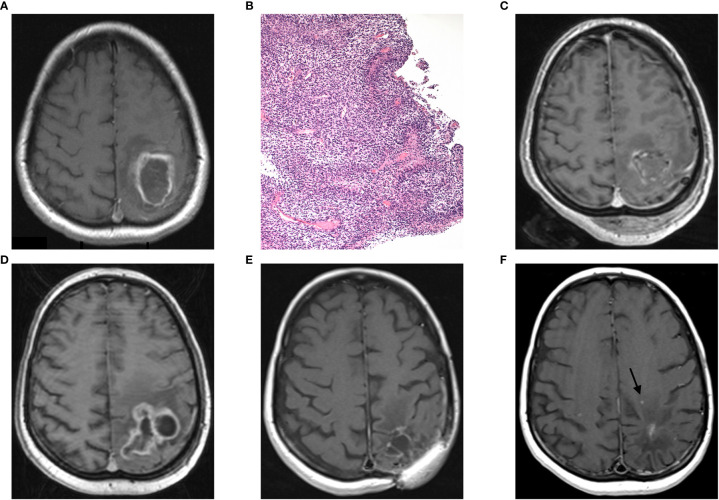
Case report 1: (A) preoperative axial MRI (postcontrast T1 weighted image) showing an intra-axial contrast-enhancing lesion in the left parietal lobe with perilesional edema extending to the central region, (B) histopathological finding of hypercellular pleomorphic glial neoplasia with multiple palisading necroses and glomeruloid microvascular proliferations (H&E, original magnification 100x), (C) postoperative MRI showing subtotal tumor resection, (D) GBM recurrence 5 months after primary tumor resection, (E) 2 years follow-up MRI, and (F) MRI with second tumor recurrence (arrow) 61 months after the first GBM recurrence.

