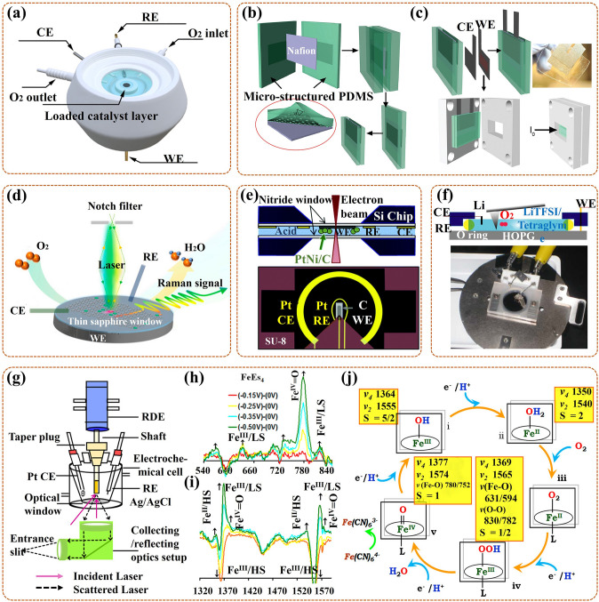
Fig. 16.
a Schematic of in-situ open XRD cell; [154] Reused with approval; Copyright 2020 Elsevier. b Fabrication schematic of the PDMS pouch, with enlarged part of the morphology for micro-structured PDMS; c Assembly schematic of in-situ XAS cell, with optical photo of the PDMS pouch [155]; Reused with approval; Copyright 2012 American Chemical Society. d Schematic of in-situ modified Raman cell [141]. Reused with approval; Copyright 2022 American Chemical Society. e Schematic of in-situ TEM liquid electrochemical chip, with a cross-sectional view of the holder for the cell on the top and the three electrodes view on the bottom [156]; Reused with approval; Copyright 2019 Royal Society of Chemistry. f Schematic of in-situ AFM cell on the top, with its optical photo on the bottom [157]; Reused with approval; Copyright 2012 American Chemical Society. g Schematic of in-situ SERRS with the RDE electrochemical cell; In-situ difference spectra of the SERRS-RDE data for Iron Porphyrin (FeEs4) h in the low-frequency extent, and i in the high-frequency extent; j Schematic of probable ORR mechanism [139]. Reused with approval; Copyright 2016 American Chemical Society

