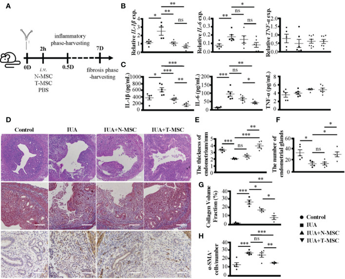
Figure 3.
TNF-α pretreatment enhances the therapeutic efficacy of MSCs in IUA mice. (A) The schematic diagram of modeling process. MSCs were stimulated by TNF-α (10 ng/ml) for 24 h to prepare T-MSCs. Both N-MSCs and T-MSCs (1 × 10⁶ cells in 200 μl of PBS) were injected through the tail vein 2 h after the operation. (B) Relative expression of mRNAs of IL-1β, IL-6, and TNF-α in murine uterine tissues. (C) The expression levels of IL-1β, IL-6, and TNF-α in serum detected by ELISA. (D) (i) The top row: H&E staining of murine uterine tissues. (ii) The middle row: Masson’s trichrome staining of murine uterine tissues. (iii) The bottom row: α-SMA protein expression detected by immunohistochemistry. (E) The statistical figure of the endometrial thickness. (F) The statistical figure of gland numbers. (G) The statistical figure of collagen volume fraction. (H) The statistical figure of numbers of α-SMA+ cells. Bar = 0.1 mm. The measurement data are presented as the means ± SEM, n = 6; *p < 0.05, **p < 0.01, ***p < 0.001. TNF-α, tumor necrosis factor-α; MSCs, mesenchymal stem cells; IUA, intrauterine adhesion; T-MSCs, tumor necrosis factor-α-primed MSCs; N-MSCs, naïve MSCs; PBS, phosphate-buffered saline. ns, no significance.

