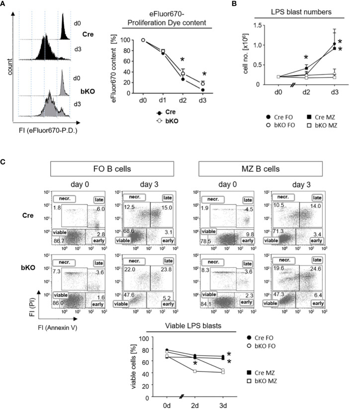
Figure 4.
DGCR8-deficiency affects the proliferation and viability of follicular and marginal zone B cells in vitro. (A) Naive splenic B cells were isolated from Cre control and DGCR8-bKO mice by magnetic cell sorting (EasySep©) and analyzed for lipopolysaccharide (LPS)-induced proliferation in vitro. Cells were stained with the proliferation dye (P. D.) eFluor670 right after isolation, and the content of the dye was measured for three days using flow cytometry. The representative figure shows eFluor670 content (day 0 and day 3). The graph depicts changes in eFluor670 fluorescence intensity of Cre and DGCR8-bKO cells normalized to the basic value determined on day 0. Points represent the mean from n=4 mice per genotype. (B) Fluorescence-based flow cytometry-sorted splenic MZ B cells (CD19+CD23lowCD21+) and FO B cells (CD19+CD23+CD21low) from Cre control and DGCR8-bKO mice were stimulated in vitro with LPS. Cell numbers were determined using flow count beads. Points represent the mean from n=3 mice per genotype. (C) Cell viability in the samples described in (B) was analyzed at different time points by flow cytometry with propidium iodide (PI) and AnnexinV. AnnexinV- and PI-negative cells were defined as viable. Mann-Whitney test was used for statistical analysis. * p<0.05; **p<0.01.

