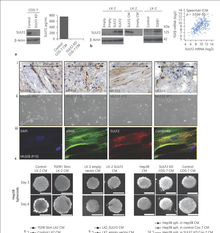
Fig. 2.
SULF2 is expressed by CAFS isolated from SULF2 stromal positive tumours, with SULF2 conditioned media stimulating growth of 3D Hep3B spheroids in vitro. a SULF2 was highly expressed in COS-7 cells and suppressed by SULF2 shRNA, assessed by Western blot and ELISA assay. b Western blot of LX-2 whole cell lysates showing LX-2 cells with little SULF2 (left panel). SULF2 expression was induced in LX-2 after transfection with a SULF2 expression vector compared to empty vector control (left). SULF2 levels were elevated in CM from LX-2 cells transfected with a SULF2 expression vector (middle). TGFβ stimulation of LX-2 cells induced SULF2 expression, with a highly significant correlation between mRNA of the two in TCGA dataset (right panels). Mixed cell isolations from SULF2 stromal positive tumours (ci) yielded CAFs after trypsinisation and replating in fibroblast culture media (cii). ciiiDual labelling immunofluorescence confirmed co-expression of SULF2 (red) and αSMA (green) in primary CAFs. CM from TGFβ-stimulated (d) or SULF2 expression vector-transfected LX-2 cells (e) promoted growth of Hep3B (SULF2 null) spheroids. f CM from control COS-7 cells expressing SULF2 promoted spheroid growth in Hep3B cells, as compared to culture with in SULF2 KD COS-7 CM. Scale bars represent 200 microns. Change in spheroid volume is in online supplementary Table 4. Data mean ± SEM; n = 7 to 10 spheroids per condition. **p < 0.01; ***p = 0.001; ****p < 0.001. “P” denotes number of passages for CAFs.

