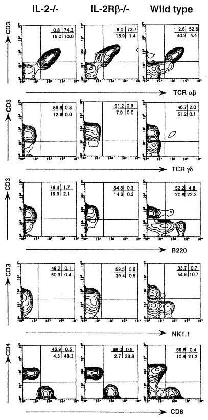
FIG. 2.
FCM analysis of the nonadherent PEC from IL-2/IL-15Rβ- or IL-2-deficient mice or wild-type mice on day 6 after i.p. infection with serovar Choleraesuis (2 × 10⁶ CFU). The cells were stained with a fluorescein isothiocyanate (FITC)-CD3 monoclonal antibody (MAb) (145-2C11) and a biotin-NK1.1 MAb (PK136) followed by streptavidin-RED 613, with FITC-CD3 (145-2C11) and a phycoerythrin (PE)–anti-TCR αβ MAb (H57-597), a PE–anti-TCR γδ MAb (GL3), or an anti-CD45R/B220 MAb (RA3-6B2) and analyzed by FCM, with the analysis gate set for small lymphocytes. In some experiments, the cells were stained with a FITC-CD3 MAb (145-2C11), a PE–anti-CD8 MAb (53-6.7), and a RED–anti-CD4 MAb (RM4-5), and the analysis gate was set for CD3+ cells. Data are representative of two independent experiments using pooled cells from three to five deficient mice and are shown as typical two-color profiles.

