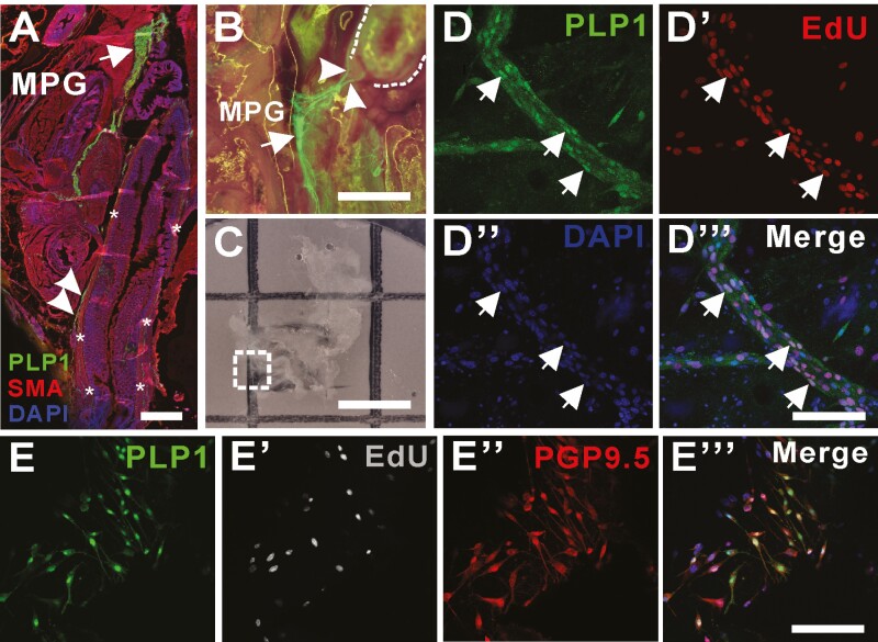
Figure 2.
PLP1-GFP glial cells residing in the extrinsic nerve fibers have a capacity to proliferate in vivo. (A) Sagittal section of pelvic cavity of Plp1-GFP mouse including distal colon (asterisks) with immunostaining with SMA shows left major pelvic ganglia (MPG, A and B, arrows) contain PLP1 expressing glial cells and fibers (A, B, arrowheads) that extends to the distal colon (A, asterisks and B, dotted line). Dissection and staining (C, dotted box) of MPG from Plp1-GFP mouse in which EdU has been administered intraperitoneally demonstrate proliferative capacity of GFP positive cells in the extrinsic colonic fibers (D-Dʹʹʹ, arrows). MPG is dissected from Plp1-GFP mouse for ex vivo organ culture for up to 7 days allowing evaluation of their capacity to proliferate (Eʹ, EdU) and to differentiate into neurons (Eʹʹ, PGP9.5) in vitro. Scale bars 500 μm (A), 200 μm (B, C), 100 μm (D-Dʹʹʹ). 50 μm (E-Eʹʹʹ), EdU, 5-ethynyl-2ʹ-deoxyuridine. Abbreviations: MPG, major pelvic ganglia; SMA, smooth muscle actin.

