Figure 3.
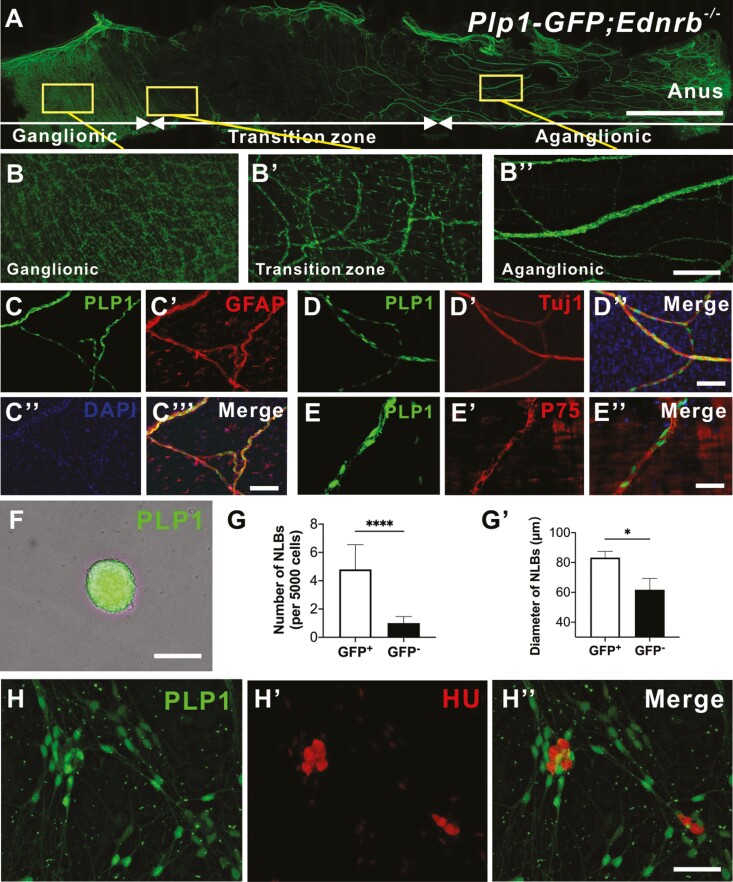
PLP1-GFP Schwann cells residing in hypertrophic nerve fibers in HSCR mice can proliferate and differentiate into neurons in vitro. Representative image of colon from Plp1-GFP;Ednrb−−- mouse (A) with high power images of ganglionic (B), transition zone (Bʹ), and aganglionic segment (Bʹʹ’). Wholemount staining of extrinsic fibers in the agangionic colon of the Plp1-GFP;Ednrb−−- mouse with GFAP (C-Cʹʹʹ), Tuj1 (D-Dʹʹ) and P75 (E-Eʹʹ). Hypertrophic fibers in the aganglionic segment of Plp1-GFP;Ednrb−/− mice were mechanically separated, sorted and cultured to form GFP positive NLBs (F). GFP positive cells generated significantly more (G, n = 5, ****P < .0001) and larger NLBs (Gʹ, n = 5, * P < .05) compared to GFP negative fraction. PLP1-GFP cells are able to generate neurons in vitro confirmed by immunoreactivity for Hu (H-Hʹʹ). Data are represented as mean ± standard deviation from 5 mice per group by Student’s t test. Scale bars 1 cm (A), 50 μm (B), 100 μm (C-F, H-Hʹʹ).

