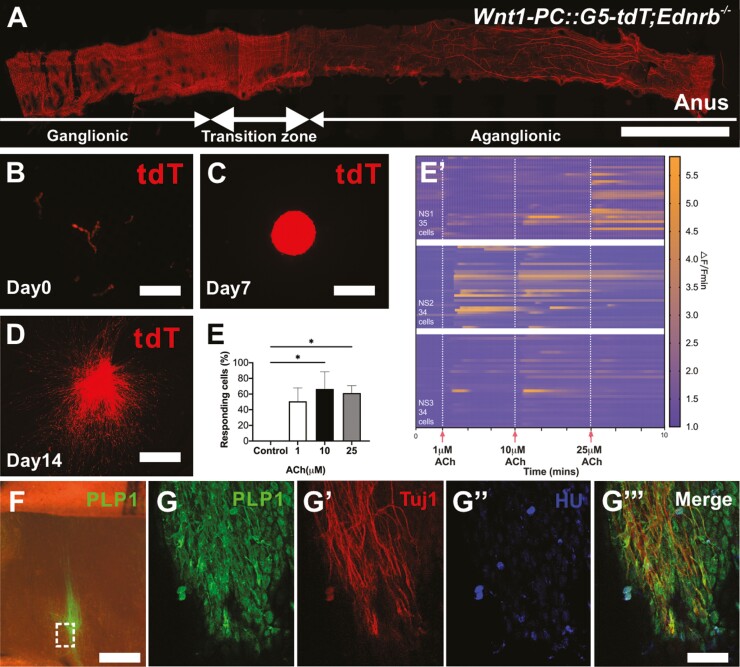
Figure 4.
Neurons generated from extrinsic nerve fibers of the aganglionic region exhibit Ca2+ activity in vitro and can proliferate and differentiate into neurons following transplantation to aganglionic mouse colon ex vivo. (A) Representative image of colon from Wnt1-PC::G5-tdT;Ednrb−/− mouse. Hypertrophic fibers in the aganglionic segment are separated mechanically (B) and cultured to form tdT+ NLBs (C). Following 14-day culture on fibronectin, prominent cell migration with extensive fiber projections are seen (D). (E) Quantification of the percentage of GCaMP5 positive cells that respond under control (unstimulated) conditions, and with 1 μM, 10 μM or 25 μM ACh (n = 3, *P < .05). (Eʹ) Calcium dynamics of cells with different concentrations of ACh (1, 10, and 25 μM, vertical white dashed lines). Data are represented as mean ± standard deviation from 34 to 35 cells per group with one-way analysis of variance. Changes in GCaMP5 fluorescence are represented on a colorimetric scale. PLP1 positive Schwann cells from hypertrophic nerves in HSCR mice are transplanted to aganglionic colon of HSCR mouse that is grown in culture ex vivo where extensive cell migration and fiber projections are seen (F). Immunohistochemistry demonstrates their neuronal differentiation in wholemount preparation (G, dotted box, G-Gʹʹʹ, arrows). Scale bars, 1cm (A), 100 μm (B-D, F), and 200 μm (G-Gʹʹʹ).

