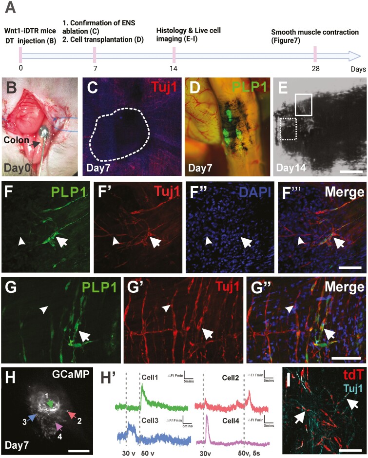
Figure 5.
Hypertrophic nerves-derived PLP1-GFP Schwann cells (HSCR-SCs) engrafted and differentiated into functional neurons following transplantation to the aganglionic colon in vivo. (A) Experimental overview using Wnt1-iDTR mouse in which all ENS express receptors for diphtheria toxin (DT). (B) Focal injection of DT with India ink to mid-colon of Wnt1-iDTR mouse (arrow) on day 0 to create colonic aganglionosis (C, dotted line). One week after DT injection (A, day 7), hypertrophic nerves-derived PLP1-GFP Schwann cells (HSCR-SCs) are transplanted to aganglionic region indicated by previous injection of India ink (D) and their successful engraftment is confirmed by histological examination 7 days after transplantation (A, day 14 and E-Gʹʹ’’). Subpopulation of transplanted cells (E, dotted line box) give rise to Tuj1+ neurons (F-Fʹʹʹ, arrows) and GFP- Tuj1+ endogenous neuronal fibers are also seen peripheral to the ablated area (F-Gʹʹ, arrowheads). High power image of cell transplantation site (E, line box) shows integration of endogenous and transplanted cell-derived neurons (G-Gʹʹ, arrows). Live cell imaging on transplanted Wnt1-GCaMP5-tdT cells isolated from hypertrophic nerves is performed 7 days following transplantation (A, day 14 and H) demonstrates variable responses to the EFS stimulation (Hʹ). Immunohistochemical staining of whole mount recipient colon shows successful ablation of endogenous ENS where transplanted Wnt1-tdT cells are engrafted (I, white dotted line) and differentiated into neurons in vivo (Iʹ Tuj1, arrows). Scale bars, 100 μm (C, E, H, I), 200 μm (F-Gʹʹ, Iʹ).

