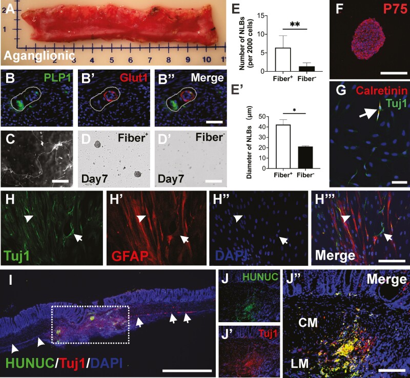
Figure 7.
Isolation and characterization of human HSCR-SCs and their transplantation to colonic aganglionosis in vivo. (A) Aganglionic colonic segment surgically resected from 6-month-old boy with HSCR. Glut1 positive extrinsic-derived nerve fibers contain PLP1-expressing cells. (B-Bʹʹ). Extrinsic fibers are separated (C), dissociated and plated for further culture. The cells from these extrinsic fibers generate more (D, E, n = 4, **P < .01) and larger (Dʹ, Eʹ, n = 4, *P < .05) NLBs than non-extrinsic fiber-derived cells. Immunohistochemical characterization of human NLBs demonstrates these NLBs contain P75+ neural crest-derived cells (F). Culturing these NLBs on fibronectin showed their capacity to differentiate into Tuj1+ neurons (G and H, arrows) and glial cells (H-Hʹʹʹ, arrowheads) in vitro. Subpopulation of differentiated neurons is also immunoreactive for calretinin (G, arrows), an enteric neuron subtype. Seven days following transplantation of these human NLBs to aganglionic mouse colon in vivo, staining for nuclei marker (I, HUNUC) identified transplanted human HSCR-SCs within the aganglionic region (I, dotted box). Immunohistochemical staining shows successful ablation of human enteric neurons (I, arrowheads) and endogenous myenteric neurons in non-ablated area of recipient colon (I, arrows) and neuronal differentiation of transplanted human HSCR-SCs (Jʹ-Jʹʹ, Tuj1, red) in vivo. Data are represented as mean ± SD per group by Student’s t-test. Scale bars 1 mm (I), 100 μm (B-Bʹʹ, D-Dʹ, G, H-Hʹʹ, J-Jʹʹ), 200 μm (C, F). Abbreviations: CM, circular muscle; LM, longitudinal muscle.

