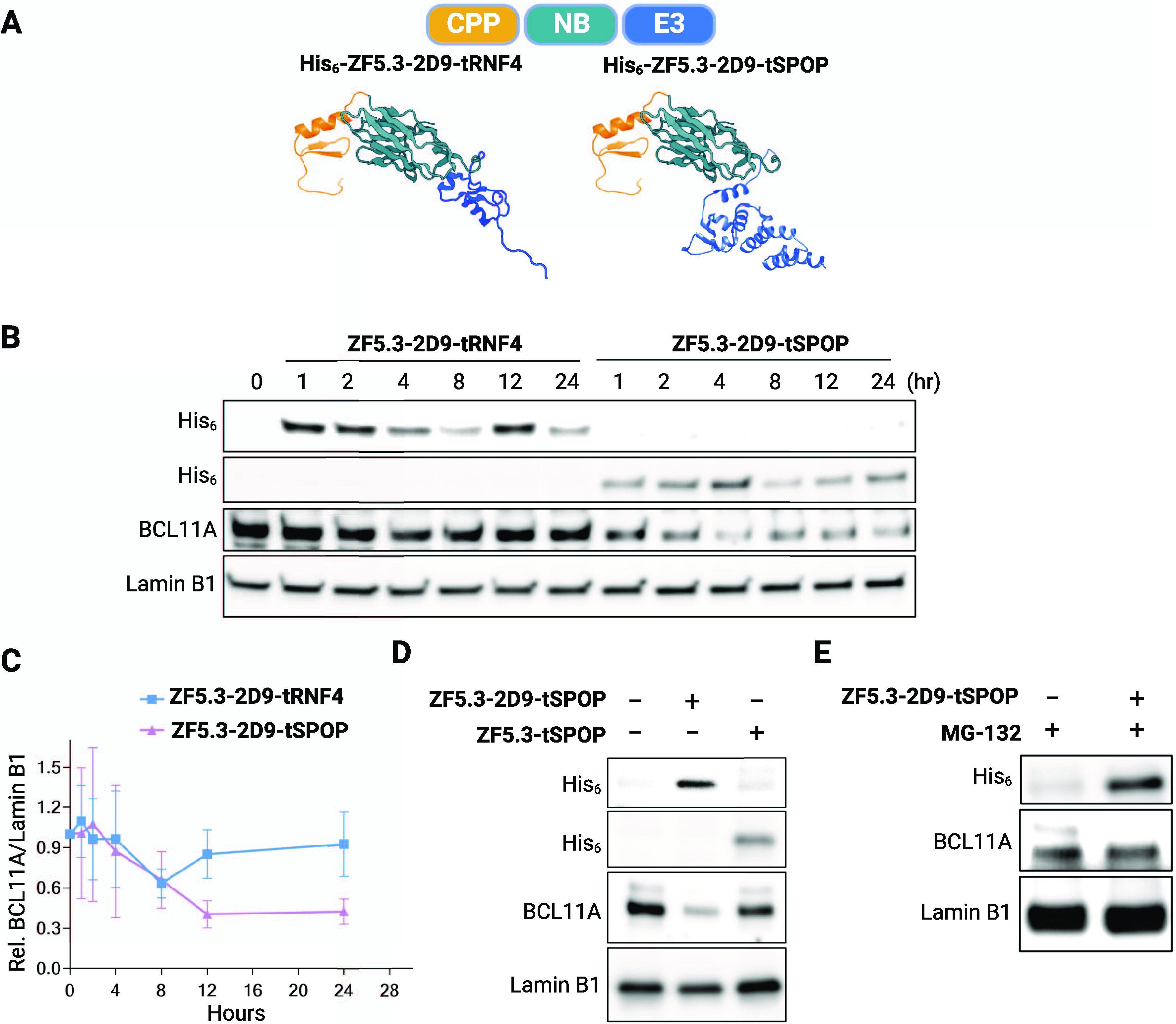
Figure 4.

Nanobody-mediated degradation of BCL11A in HUDEP-2 cells. (A) Schematic depiction of BCL11A degraders. The individual structures of the RNF4 RING domain (4PPE), the BTB domain of SPOP (3HTM), ZF5.3 (modeled secondary structure), and 2D9 (7UTG) were retrieved from the Protein Data Bank. (B) Representative immunoblot showing loss of BCL11A in HUDEP-2 cells treated with ZF5.3-2D9-tRNF4 or ZF5.3-2D9-tSPOP. (C) Quantification of BCL11A loss by immunoblots (mean ± SD, n = 3). Immunoblots revealing that (D) loss of BCL11A requires the presence of 2D9 and (E) is prevented by addition of 5 μM of the proteasome inhibitor MG-132. Lamin B1 was used as a loading control for the immunoblots.
