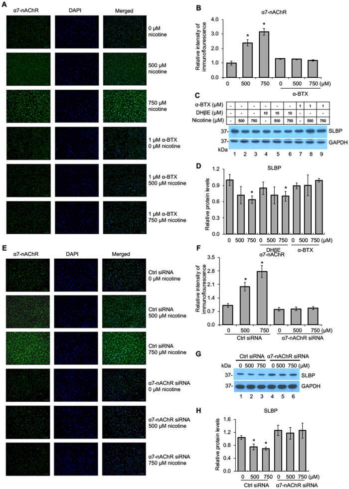
Figure 2.
Nicotine-mediated downregulation of SLBP is α7-nAChR dependent. A–B, Immunofluorescence (IF) staining of α7-nAChR in BEAS-2B cells treated with or without nicotine for 24 h in the presence or absence of 1 μM α-BTX, an inhibitor of α7-nAChR. Representative IF staining for α7-nAChR, DAPI, and merged images are shown (A). The IF intensities for α7-nAChR were determined by ImageJ software, normalized to control group, and presented as bar graphs (B). C and D, α-BTX, an inhibitor of α7-nAChR, but not DHβE, an inhibitor of α3/4-nAChR, attenuates nicotine-induced downregulation of SLBP. BEAS-2B cells were treated with or without nicotine for 24 h in the presence or absence of DHβE or α-BTX and subjected to Western blot analysis with indicated antibodies (C). The band intensities were quantified and presented as bar graphs to show relative protein levels for SLBP (D). GAPDH was used as an internal control. E–F, IF staining of α7-nAChR following nicotine treatment of BEAS-2B cells transiently transfected with the control siRNA or the siRNA specific for α7-nAChR. Representative IF staining for α7-nAChR (green), DAPI (blue), and merged images are shown (E). The IF intensities for α7-nAChR were determined by ImageJ software, normalized to control group, and presented as bar graphs (F). G and H, Knockdown of α7-nAChR by siRNA attenuates nicotine-mediated downregulation of SLBP. BEAS-2B cells that have been transiently transfected with the control siRNA or the siRNA specific for α7-nAChR were treated with or without nicotine for 24 h and subjected to Western blot analysis with indicated antibodies (G). The band intensities were quantified and presented as bar graphs to show relative protein levels for SLBP (H). GAPDH was used as an internal control. Untreated controls in lane 1 were used as references, which were set to 1. The data shown are the mean ± SD (n = 3). *p < .05 versus untreated control group.

