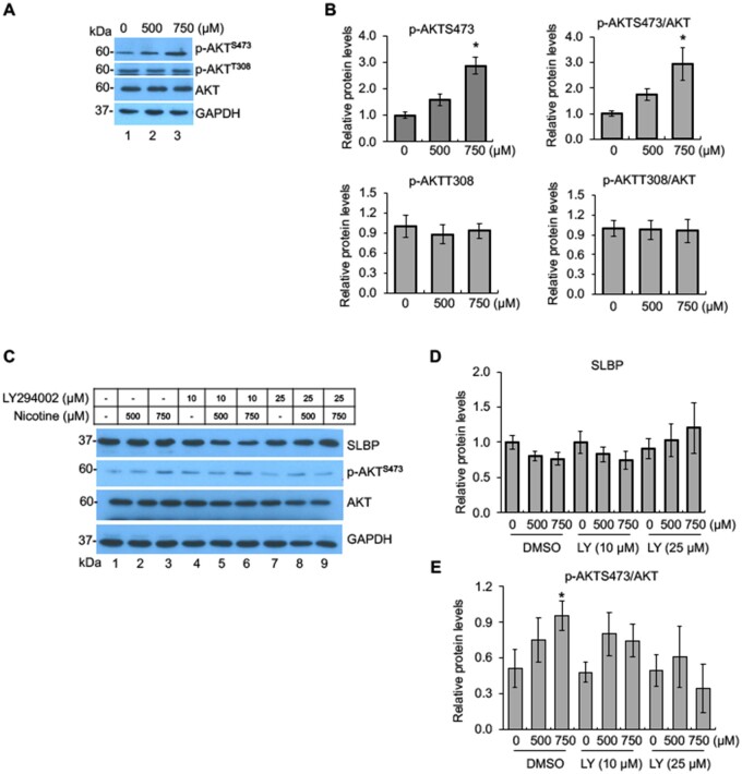
Figure 3.
Activation of PI3K/AKT signal transduction pathway by nicotine exposure. A and B, Phosphorylation of AKT at S473 by nicotine. BEAS-2B cells were treated with or without nicotine for 24 h followed by Western blot analysis with indicated antibodies (A). The band intensities were quantified and presented as bar graphs (B). GAPDH was used as an internal control. C–E, Inhibition of PI3K/AKT pathway attenuates nicotine-induced downregulation of SLBP. BEAS-2B cells were treated with or without nicotine for 24 h in the presence or absence of LY294002, an inhibitor of PI3K, and subjected to Western blot with indicated antibodies (C). The band intensities were quantified and presented as bar graphs to show relative quantifications of SLBP (D) and p-AKTS473/AKT (E). GAPDH was used as an internal control. Untreated controls in lane 1 were used as references, which were set to 1. The data shown are the mean ± SD (n = 3). *p < .05 versus untreated control group.

