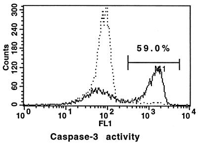
FIG. 7.
Comparison of caspase-3 activities of resting monocytes and monocytes after phagocytosis of S. aureus. The cells were cultured for 2 h with (solid line) or without (dotted line) bacteria, loaded with a caspase-3 fluorogenic substrate, and incubated for another hour. Intensity of green fluorescence (x axis), corresponding to caspase-3 activity, is shown.

