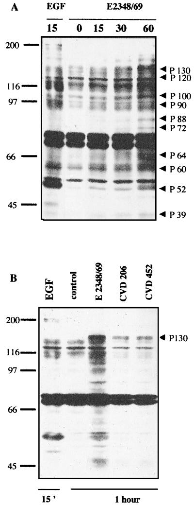
FIG. 1.
Induction of protein tyrosine phosphorylation in T84 cells infected with EPEC. (A) T84 cells were lysed at various times after infection. Samples were resolved by electrophoresis on an SDS–9% polyacrylamide gel and analyzed by immunoblotting using antiphosphotyrosine antibody. In both panels, the positions of molecular weight standards are shown at the left in kilodaltons. The putative T84 cell-induced tyrosine-phosphorylated proteins are indicated by arrowheads. (B) T84 cells were infected for 1 h with WT strain E2348/69 and mutant strains CVD206 and CVD452. Samples were resolved as described above. The mutant-induced tyrosine phosphorylated protein of 130 kDa is indicated by an arrowhead. The control lane corresponded to uninfected cells. The positive control lane was obtained from cells treated with EGF (10 nM, 15 min). Identical results were obtained at least five times.

