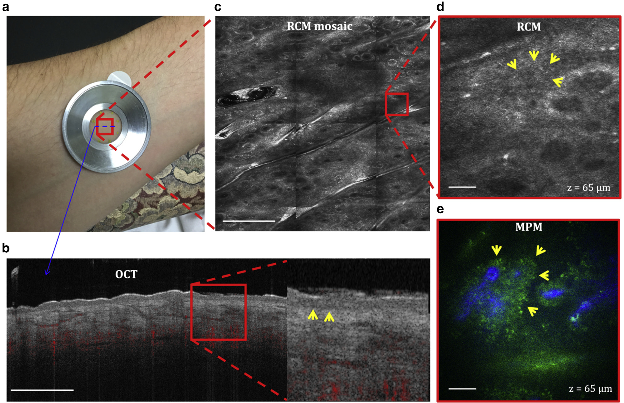
Figure 2. Normal skin.

(a) Imaging location on the volar forearm of a volunteer. All the three devices use a metallic ring that attaches to the skin by double-sided tape and to the instrument imaging head magnetically to ensure image stability. (b) Cross-sectional (B-scan) OCT image of the skin showing the ability to capture the dermal vascular network (red) and to differentiate the epidermal from the dermal layer by clearly identifying the basal membrane (arrows in the close-up image). Bar = 1 mm. (c) Enface (horizontal) RCM mosaic (mm-scale) image showing the overall morphology of the skin at a depth of 65 μm (DEJ). Bar = 500 μm. (d) Close-up view of the area outlined in the RCM image in c revealing the keratinocytes of the basal layer surrounding the dermal papilla (arrows). Bar = 40 μm. (e) Enface MPM image acquired at the same depth showing a similar morphology with the RCM image but with enhanced contrast that clearly distinguishes the dermal papilla collagen (blue) from the surrounding keratinocytes (green, arrows). Bar = 40 μm. DEJ, dermal–epidermal junction; MPM, multiphoton microscopy; OCT, optical coherence tomography; RCM, reflectance confocal microscopy.
