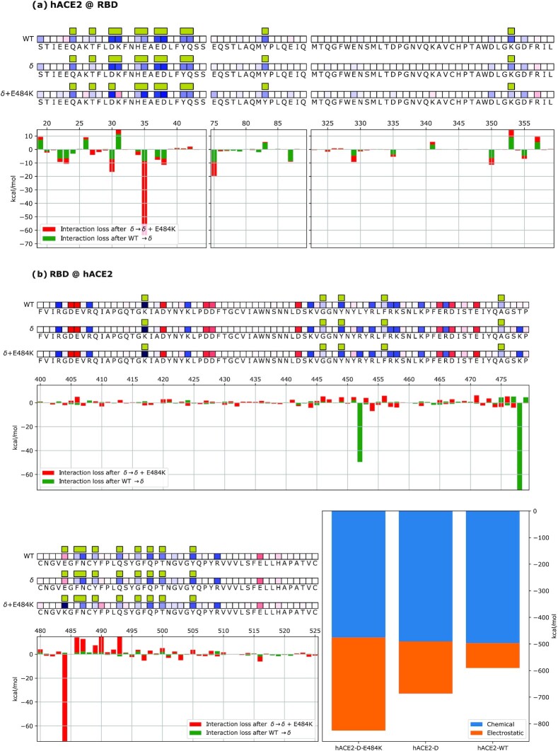
Fig. 5.
Mechanistic characterization of spike–hACE2 binding suggests that Delta + E484K spike has stronger hACE2 binding than the Delta variant. (a) Data are plotted on hACE2 primary structure bound to the Wuhan spike (WT), Delta spike (δ) and Delta + 484 K spike (δ + 484 K). Amino acids are represented by the corresponding letters and numbered on the histogram's horizontal axis. Interface residues are highlighted by yellow bars and their overall effect on the other molecule is indicated by red (repulsive) or blue (attractive) squares (energy scale is identical to the one employed in the other figures). Histograms underneath the sequences show the relative change in binding energy (green: Delta compared to Wuhan; red: Delta + E484K compared to Delta). (b) Data are plotted on the viral spike primary structure bound to the Wuhan spike (WT), Delta spike (δ), and Delta + 484 K spike (δ + 484 K). Amino acids are represented by the corresponding letters and numbered on the histogram's horizontal axis. Interface residues are highlighted by yellow bars and their overall effect on the other molecule is indicated by red (repulsive) or blue (attractive) squares (energy scale is identical to the one employed in the other figures). Histograms underneath the sequences show the relative change in binding energy (green: Delta compared to Wuhan; red: Delta + E484K compared to Delta). Bar plots on the bottom right represent the overall binding energy of hACE2 with the Wuhan, Delta, and Delta + E484K strains, partitioned into chemical or electrostatic contributions.

