Fig. 5.
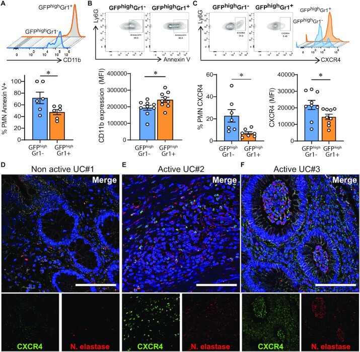
Different PMN dynamics correlates with different CXCR4 expression and apoptosis. Lamina propria PMNs were isolated 6 h post-wounding from mice subjected to DSS for 4 days. PMNs were identified by FACS as CD45+CD11b+Ly6G+. Two populations were distinguished based the presence or absence of Gr1+ staining. (A to C) Representative plots and quantification of expression of (A) CD11b, (B) apoptosis as determined by Annexin V binding, and (C) CXCR4 in GFPhighGr1+ and GFPhighGr1− lamina propria PMNs from mice subjected to DSS + wounding. Data are mean ± SEM of two independent experiments (n = 5 to 7 mice per group; *P < 0.05; **P < 0.01 by two-tailed Student’s t-test). (D to F) Immunofluorescence staining of CXCR4 (green), Neutrophil elastase (red), and nuclei (blue) in colonic mucosa biopsies from patients with IBD. Representative confocal micrographs of (D) nonactive UC and (E and F) active UC. Scale bars: 100 μm.

