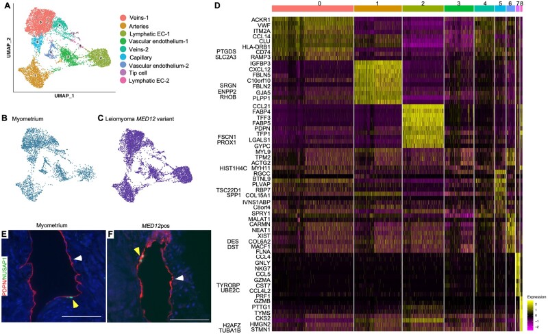
Figure 4.
Lymphatic ECs are present in leiomyomas. (A) Uniform Manifold Approximation and Projection on Principal (UMAP) components showing the clusters of 10 448 endothelial cells. (B, C) UMAP showing the cluster annotation per condition: myometrium and MED12-variant positive leiomyomas. (D) Heatmap of endothelial cell clusters. The colored bar on the top represents cluster number. Columns denote cells; rows denote genes. (E, F) Immunostaining for PDPN (white arrowheads) and NUSAP1 (yellow arrowheads) showing the presence of Lymphatic EC clusters in normal myometrium (E) and MED12-variant positive leiomyomas (F). Scale bar is 100 µm.

