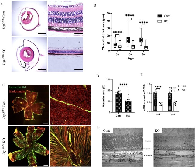
Fig. 2.
Decrease of Vegf expression from RPEs and abnormal development of choriocapillaris are observed in Lrp2RPE KO mice. (A) The chordal region in the representative H&E-stained cross-sections of enucleated eyes from 6-week-old Lrp2RPE Cont and Lrp2RPE KO mouse showing enlarged overall eye size and thinner choroidal thickness in KO mouse (bottom). Scale bar: 1 mm (left panels), 50 µm (right panels). (B) Choroidal thickness measurements in 3-, 6-, and 8-week-old Lrp2RPE Cont and Lrp2RPE KO mouse showing reduction of choroidal thickness in KO mouse. (C) Representative immunohistochemistry of choroid flat-mount (green: isolectin B4, red: endomucin) and (D) quantification of vascular area in choroid of 8-week-old Lrp2RPE Cont and Lrp2RPE KO mouse showing reduction of choriocapillaris in KO mouse. Scale bar: 1 mm (left panels), 20 µm (right panels). (E) Transmission electron microscope observation of choroids of 8-week-old Lrp2RPE Cont and Lrp2RPE KO mouse. Choriocapillaris highlighted by dashed line in Cont (left) is disappeared in KO (right), resulting in choroidal thinning. Scale bar: 100 µm. (F) Lrp2 and Vegf mRNA expression from primary RPEs in 6-week-old Lrp2RPE Cont and Lrp2RPE KO mouse. Graphs represent as mean ± SD. n = 5. ***P < 0.001, ****P < 0.0001, two-tailed Student's t-tests.

