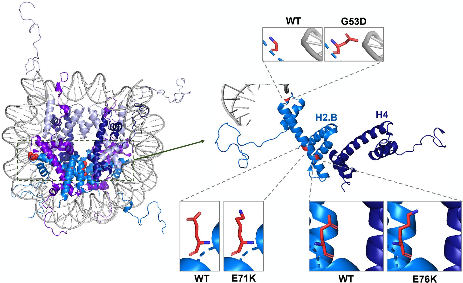
Figure 3:

Nucleosome crystal structure with non-canonical histone mutations highlighted in red (Left). (Right) Close up of Histone H2.B (aqua blue) interacting with DNA (Gray) and H4 (Dark Blue). Side by side images of WT and mutated amino acid at reside 53, 71, and 76 (PDB code 1KX5).
