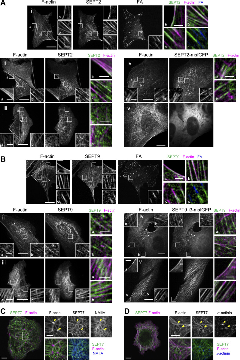
Figure S1.
SEPT2, SEPT7 and SEPT9 distribution on different types of stress fibers in U2OS cells. (A) Representative confocal micrographs of SEPT2 immunostained cells (i–iii) and cells expressing SEPT2-msfGFP (iv and v). SEPT2 immunostained cells are co-stained for F-actin (phalloidin) and the FA protein paxillin. Examples show SEPT2 localizing (i) to peripheral (a) and ventral (b and c) SFs and excluded from focal adhesions (FAc) , (ii) to peripheral (a) and perinuclear actin caps (b), (iii) to transverse arcs (b) and excluded from dorsal SFs (a and b), (iv) to transverse arcs (a and b) and excluded from dorsal SFs (a), and (v) showing a diffuse cytosolic phenotype. (B) Representative confocal micrographs of SEPT9 immunostained cells (i–iii) and cells expressing SEPT9_i3-msfGFP (iv and v). SEPT9 immunostained cells are co-stained for F-actin (phalloidin) and the FA protein paxillin. Examples show SEPT9 localizing (i) to peripheral (a) and ventral (b and c) SFs and excluded from focal adhesions (FA; c), (ii) to perinuclear actin caps (a and b), (iii) to transverse arcs (a) and ventral SFs (b), (iv) to transverse arcs (a) and excluded from dorsal SFs (a) and to ventral SFs (b), and (v) to peripheral (a) and perinuclear actin caps (b). (C and D) Representative confocal micrographs of SEPT7 immunostainings showing SEPT7 localizing to ventral actin nodes. Cells are co-stained for F-actin (phalloidin) and non-muscle myosin heavy chain IIA (NMIIA; C) or α-actinin (D). Yellow arrowheads point to two actin nodes in each example. Scale bars in large fields of views, 10 μm. Scale bars in insets, 2 μm (A and B) and 5 μm (C and D). Related to Fig. 1 B.

