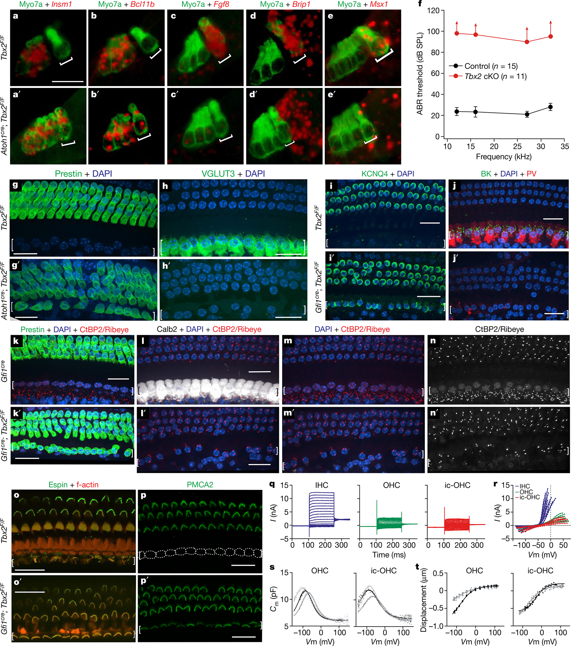
Fig. 1 |. Early ablation of Tbx2 results in the generation of OHCs in the position of IHCs.

a–e′, In situ hybridizations (red) with immunohistochemistry for hair cells using myosin VIIa (green) on E17.5 cochleae after conditional Tbx2 deletion. In mutants, embryonic hair cells in the position of IHCs (square brackets) express markers of developing OHCs (a–b′) but not those of IHCs (c–e′). Expression of Brip1 and Msx1 in supporting cells near IHCs is unaltered in conditional knockouts (cKOs) (n ≥ 3 biologically independent samples). f, ABRs are absent (up-facing arrows) from mature or weaned (P26–P39) mice with embryonic ablation of Tbx2 (red; n = 11: 5 Atoh1cre/+; Tbx2F/F plus 6 Gfi1cre/+; Tbx2F/F). Littermate controls (black; n = 15: Tbx2F/F, Atoh1cre/+; Tbx2F/+, Gfi1cre/+; Tbx2F/+ and Gfi1cre/+) had normal ABRs. Error bars, s.d. g–p′, Immunohistochemistry of mature cochleae indicating that, after embryonic Tbx2 ablation, all hair cells in the position of IHCs (square brackets) displayed all examined features of OHCs but none for IHCs. These features include expression of prestin (g, g′, k, k′), KCNQ4 (i, i′) and PMCA2 (p, p′); lack of VGLUT3 (h, h′), BK, parvalbumin (j, j′), CALB2 (l, l′) and nuclear CtBP2 (m–n′); few synaptic ribbons (Ribeye+ puncta; k–n′); small nuclei (DAPI+; m, m′) located at the base and not the middle of the hair cell (h, i′, j); and shorter stereocilia tightly bundled as in OHCs rather than the long and fanning pattern for IHCs (o–p′). Dotted lines (p) delineate IHCs, which lack PMCA2 (n ≥ 3 biologically independent samples). q–t, Whole-cell currents from ic-OHCs (of Fgf8creER; Tbx2F/F; R26LSL-tdTomato/+ mice treated with tamoxifen at birth; tdTomato+) and from control IHCs and OHCs. q, r, Current responses to voltage steps (−140 mV to +80 mV) in ic-OHCs (n = 7) were characteristic of OHCs (n = 7) but not IHCs (n = 12). s, t, Electromotility assessment from nonlinear capacitance (s) and video recordings (t). Two-state Boltzmann fits. Similar to control OHCs (n = 9), ic-OHCs (n = 10) were electromotile. Hair cells originated from the apical one-fourth of the cochlea at P25–P29. The cells were from ≥3 animals. Scale bars, 20 μm.
