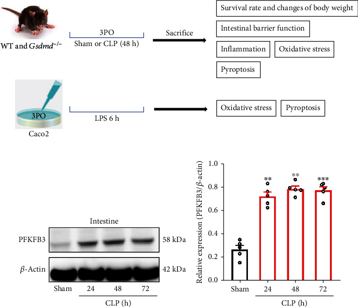
Figure 1.

The expression of PFKFB3 was elevated in the intestinal tissues of septic mice. (a) The experimental design of this study. (b, c) The expression of PFKFB3 was analyzed by Western blot at different time points after CLP. Experimental values are expressed as means ± SD (n = 5 per group). Statistical analysis was performed using one-way ANOVA (c). ∗∗P < 0.01 and ∗∗∗P < 0.001.
