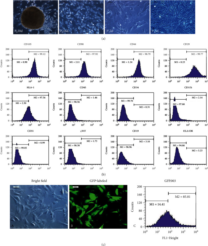
Figure 1.

The hUCMSC isolation, culture, and identification. (a) The hUCMSCs were isolated and cultured by adherent method, as well as grew in vortex shape. (b) The hUCMSCs are positive expression of CD105, CD90, CD44, and CD29 and negative expression of CD45, CD34, CD11b, CD31, vWF, CD19, and HLA-DR using flow cytometry. (c) The hUCMSCs were labeled with green fluorescent protein (GFP) using a lentiviral strategy (GFP-hUCMSCs).
