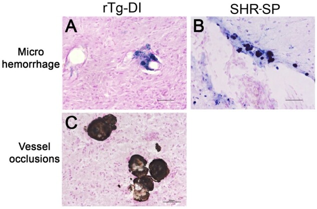
FIGURE 5.

Cerebral microhemorrhages in rTg-DI rats and SHR-SP rats. Brain sections from 12-month rTg-DI rats (A, C) and SHR-SP rats (B) were stained for hemosiderin with Prussian blue to identify microhemorrhages (blue). Scale bars = 50 µm. Representative images show that cerebral microhemorrhages in the thalamus of rTg-DI rats (A) and cortex of SHR-SP rats (B). In addition, rTg-DI rats typically show calcified, occluded small vessels detected by von Kossa calcium stain in the thalamic region (C) that are not observed in SHR-SP rats.
