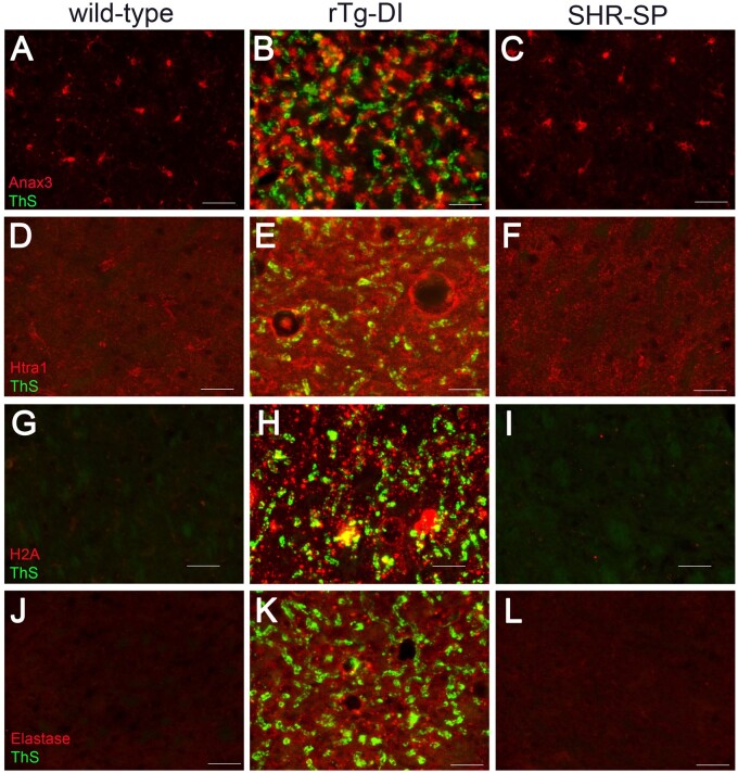
FIGURE 9.
Increased immunolabeling for specific protein markers in rTg-DI rats. Brain sections from 12-month WT rats (A, D, G, J), rTg-DI rats (B, E, H, K), and SHR-SP rats (C, F, I, L) were stained with thioflavin S to detect microvascular fibrillar amyloid (green) and rabbit polyclonal antibody to annexin 3 (A–C), Htra1 (D–F), histone 2A (G–I), and neutrophil elastase (J–L) (red). Scale bars = 50 µm. Representative images show each protein is increased only in the thalamus of rTg-DI rats.

