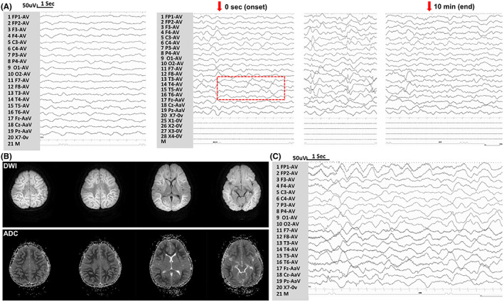
FIGURE 2.

EEG and neuroimaging results in the cases with KCNH1 variants. (A) EEG of case 1 detected diffuse slow waves, and subclinical seizures originated from bilateral temporal (red box indexed the seizure onset). (B) Brain MRI showed bright tree appearance in DWI, and low‐intense signal in ADC. (C) EEG of case 3 indicated diffuse slow waves and drug‐related fast waves. (DWI, diffuse weighted images; ADC, apparent diffusion coefficient)
