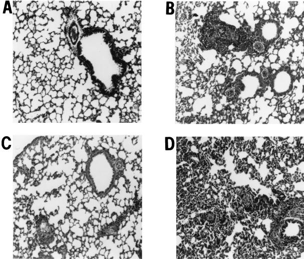
FIG. 2.
Lung histopathology in C57BL/6, IL-4−/−, and IFN-γ−/− mice. C57BL/6, IL-4−/−, and IFN-γ−/− mice were immunized and injected i.v. with 200,000 live Mf as described above. One week later, animals were sacrificed, lungs were fixed in formalin, and 5-μm sections were stained with hematoxylin and eosin. Shown are representative lung sections from similar sites in the left lobe for naïve mice (A), immunized, Mf-challenged C57BL/6 mice (B), IL-4−/− mice (C), and IFN-γ−/− mice (D). Peribronchial and perivascular cell infiltrates can be detected in panels B and D. In panel D, normal lung architecture is also lost. Sections are representative of five mice per group from two repeat experiments.

