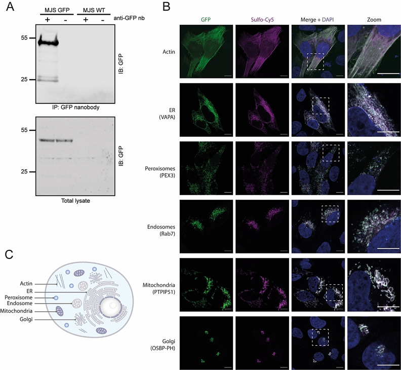
Figure 5.
A. Western blot analysis of the pull‐down of GFP‐Rab7 from cell lysate using GFP Nb‐biotin conjugate 6. The signal around 25 kDa is GFP as a result of protein degradation. B. Confocal images of MelJuSo cells expressing GFP‐Rab7 in the presence of GFP Nb‐Cy5 7. C. Illustration of a cell highlighting the cellular compartments visualized in B.

