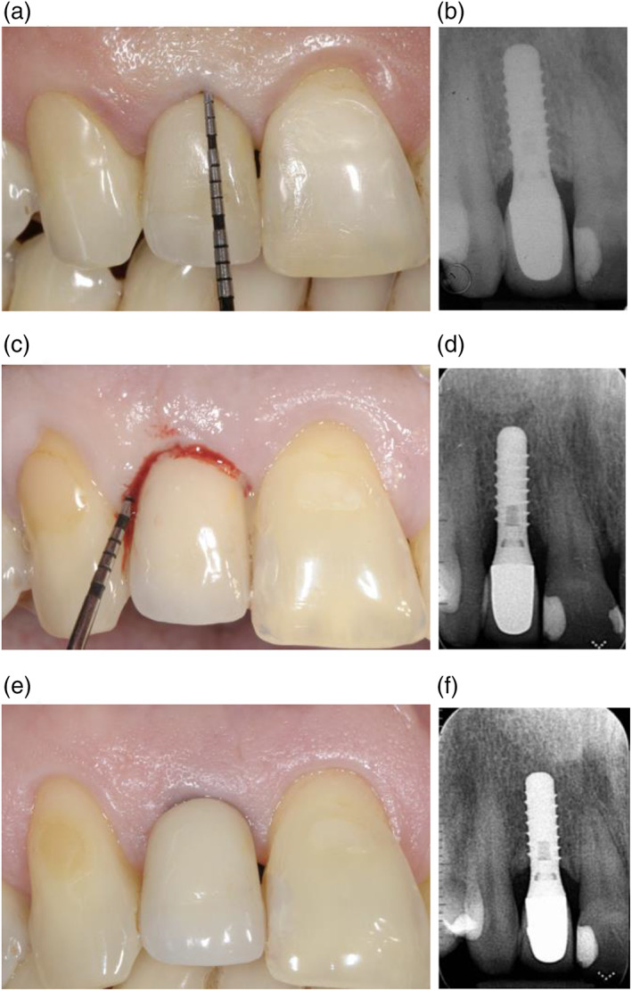
FIGURE 1.

Clinical and radiographic images of an implant placed in April 2001 in an “adherent patient” (a,b); in May 2017, the site exhibited clinical signs of inflammation, bleeding on probing, increased probing depth and radiographic bone loss (c,d); clinical and radiographic images at the 20‐year follow‐up, following surgical treatment of the biological complication (e‐f)
