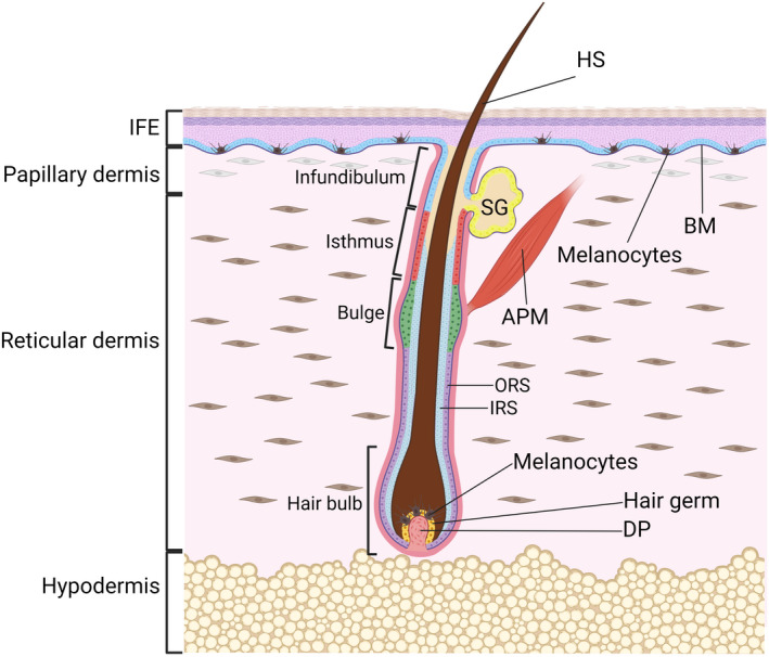
FIGURE 1.

Morphology of the skin. The epidermis and the underlying dermis are separated by a basement membrane (BM). Multiple, spatially distinct stem cell populations have been identified in the interfollicular epidermis (IFE) and the bulge, isthmus, infundibulum and sebaceous gland (SG) parts of the hair follicle and are indicated by different colours. Two populations of fibroblasts populate the dermis: papillary fibroblasts are in proximity to the BM, while reticular fibroblasts are found in the central dermis. The hair follicle is depicted in the growth phase (anagen), when a transient population of stem cells in the hair germ create an inner root sheath (IRS) and hair shaft (HS, protruding out of the skin surface), while stem cells in the permanent bulge region of the hair follicle give rise to the outer root sheath (ORS). The hair germ rests above the dermal papilla (DP), a population of mesenchymal cells that provides inductive signalling for hair growth and modulates hair follicle regeneration. Pigment‐producing melanocytes are present in the hair follicle and the IFE. APM, arrector pili muscle. Figure graphics were created with BioRender.com.
