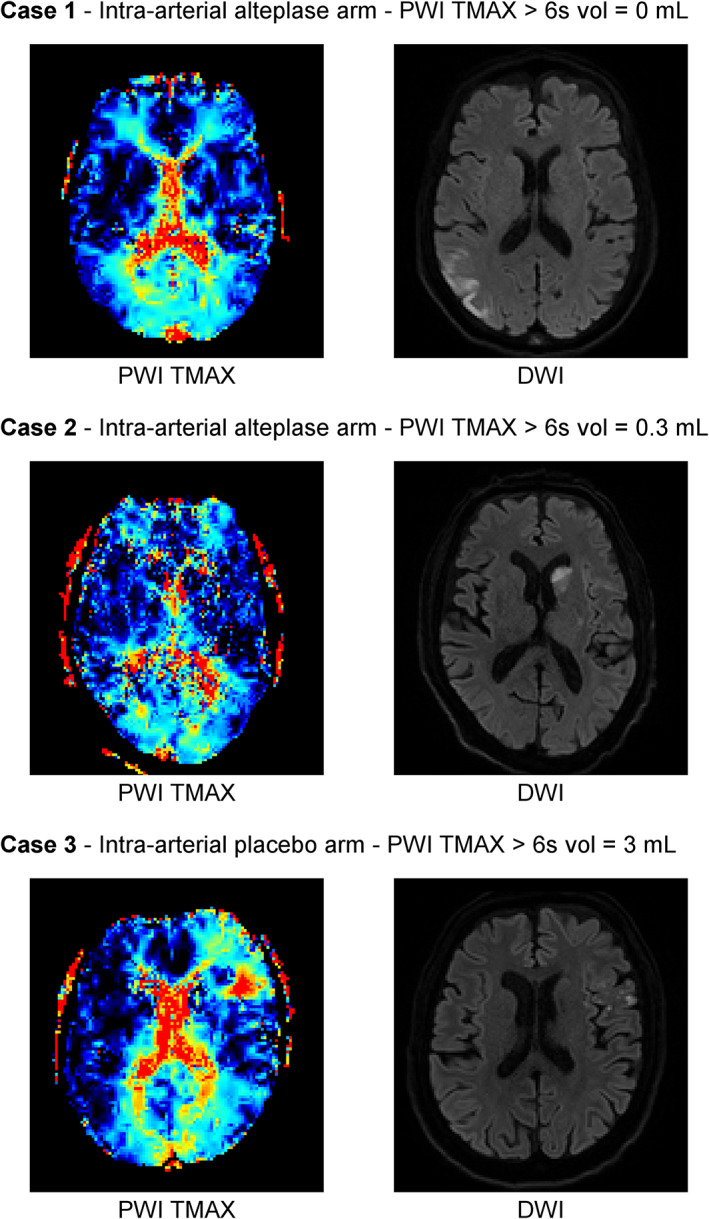
FIGURE 3.

Representative cases of perfusion patterns on follow‐up magnetic resonance imaging. DWI = diffusion‐weighted imaging; PWI = perfusion‐weighted imaging; TMAX = time to maximum.

Representative cases of perfusion patterns on follow‐up magnetic resonance imaging. DWI = diffusion‐weighted imaging; PWI = perfusion‐weighted imaging; TMAX = time to maximum.