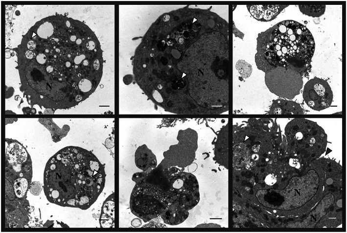
Figure 4.

Electron micrographs of chicken embryo kidney (CEK) cells infected with the IBV strain VicS‐v for 48 h. The white arrow heads indicate the location of viral particles in some of the vesicular packets. The grey arrow head indicates the location of the junction of two adjacent cells, which are forming a syncytium. N, nuclei. Scale bars represent 500 nm (white) and 1 μm (black). Photograph, courtesy of Ms Liliana Tatarczuch, Melbourne Veterinary School, Faculty of Veterinary and Agricultural Sciences, The University of Melbourne.
