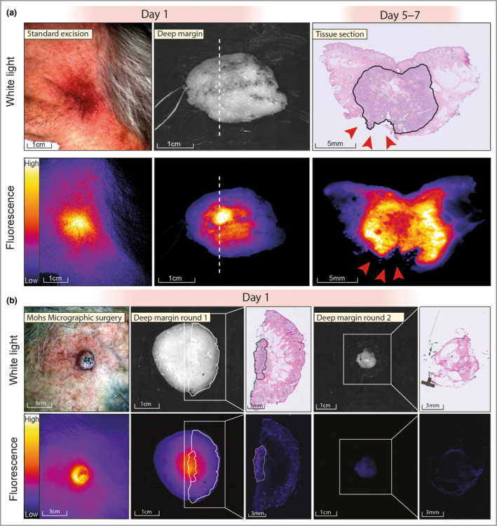
Figure 1.

Fluorescence molecular imaging during standard excision and Mohs micrographic surgery (MMS). (a) Fluorescence molecular imaging before standard excision of a temporal, subdermal tumour that was earlier irradically removed. Ex vivo imaging of the specimen showed a fluorescent lesion at the deep resection margin, correlating with a tumour‐positive margin on histopathology (red arrowheads). (b) Fluorescence molecular imaging during an MMS procedure. In vivo imaging shows a sharply demarcated fluorescent lesion. Ex vivo imaging shows a fluorescent lesion at the deep resection margin, which colocalizes with tumour on haematoxylin and eosin histopathology. The second MMS round did not show any remaining fluorescence signal, and no tumour was found on histopathology.
