
FIGURE 1.
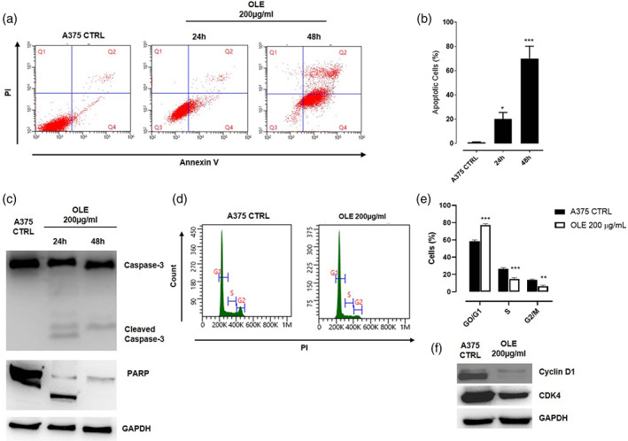
Olive leaf extract (OLE) induced apoptotic cell death and cell cycle arrest in human melanoma cells. (a‐b) Apoptotic cell death in human A375 cells was detected by annexin V/propidium iodide (PI) staining. A375 cells were treated with OLE 200 μg/ml and apoptosis was determined at 24 and 48 hr by flow cytometric analysis. The percentage of apoptotic cells are shown in the bar diagram as the mean ± SEM (n = 3), (*p < .05; ***p < .001 vs. A375 CTRL). (c) Western blot analysis of caspase‐3 and PARP in A375 whole‐cell lysates following the treatment with OLE (200 μg/ml) for 24 and 48 hr. GAPDH was detected as loading control. The blot shown are representative of three independent experiments. (d‐e) Cell cycle distribution was analyzed in human A375 cells treated with OLE (200 μg/ml) for 24 hr by flow cytometry following PI staining. Data are expressed as mean ± SEM of three independent experiment (**p < .01; ***p < .001 vs. CTRL). (f) Western blot analysis of Cyclin D1 and CDK4 carried out in A375 whole‐cell lysates following the treatment with OLE (200 μg/ml) for 24 hr. GAPDH was detected as loading control. The blot shown are representative of three independent experiments