
FIGURE 3.
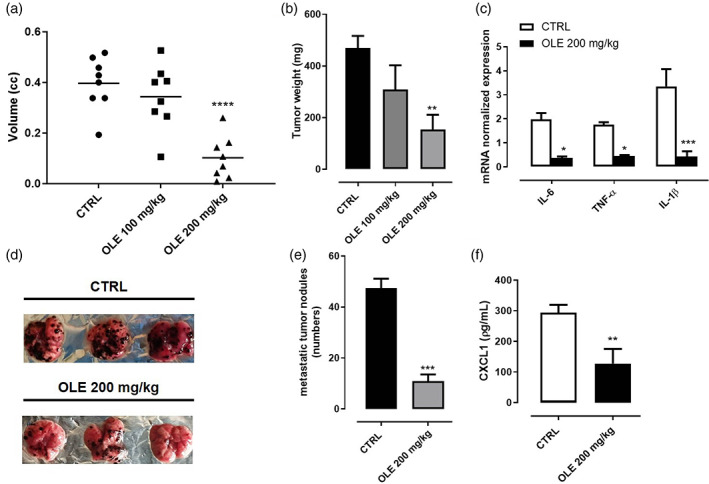
Olive leaf extract (OLE) inhibited tumor growth and suppressed the lung metastasis in B16F10‐bearing mice. B16F10 mouse melanoma cells were subcutaneously injected in the right flank of C57BL/6 mice. OLE (100 and 200 mg/kg) was given orally to mice, while control mice received vehicle only. (a‐b) Volume and weight of dissected tumor were measured after 14 days of treatment. Data are presented as mean ± SEM (n = 8). (**p < .01; ****p < .0001 vs. CTRL). (c) Expression of il‐6, tnf‐α and il‐1β assessed by qPCR analysis in dissociated tumor tissues. Data are expressed as mean ± SEM (n = 8); (*p < .05; ***p < .001 vs. CTRL). B16F10 mouse melanoma cells were injected through the lateral tail vein of C57BL/6 mice. OLE (200 mg/kg) was given orally to mice, while control mice received vehicle only. (d) Representative photographs of lung tissue excised at day 14th after treatment. (e) The metastatic nodules were counted, and data presented as the mean ± SEM (n = 8); (***p < .001 vs. CTRL). (f) Quantification of CXCL1 plasma levels assessed by ELISA in tumor bearing mice. Data are expressed as mean ± SEM (n = 8); (**p < .01 vs. CTRL)