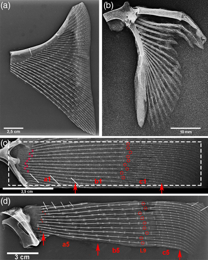
FIGURE 2.

Raja clavata (Chondrichthyes) X‐rays of specimens 1 & 5. (a) Specimen 5, high‐resolution X‐rays of the propterygium wing‐fin anterior sector (after dissection of skin and muscles) showing the increasing number of radials from the anterior tip towards the central sector and the joint angles. In the outer band (with skin not dissected), the dermal denticles are superimposed on the thin, most external radials. (b) Specimen 5, X‐rays of pelvic basipterygium with the 1st line of long and thick radials. The thicker compound radial forms a diarthrodial joint with the pelvic girdle. (c–d) X‐rays of the 9 central sectors rays of specimens 1 and 5 with the labeled references for morphometry.
