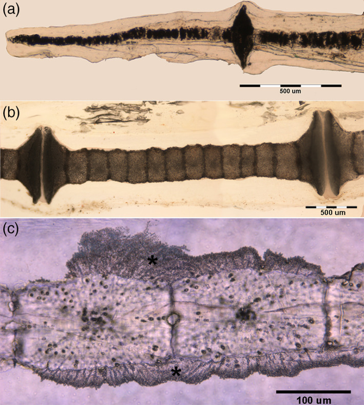
FIGURE 5.

Raja clavata (Chondrichthyes). Heat‐deproteinated (400°C) images of wing‐fin radials at different ray levels. (a) (40x) Small, globular Ca2PO4 spherules of the radials apical segment mixed with black carbon deposits produced by combustion of the organic matrix. (b) (100x) Mono‐columnar radialshowing the calcified, cylindrical tiles: at the proximal and distal ends, the mineralized cartilage enlarges to form the disks of inter‐radial joints. (c) (200x) Detail of cylindrical tiles showing a separation plane between individual tiles. The black dots in the calcified tissue mass are carbon deposits into the chondrocyte lacunae. The compact calcified mass extending externally over several neighboring tesserae corresponds to a different type of mineralization which has also been reported around polygonal tesserae of the endoskeleton as “endophytic masses”.
