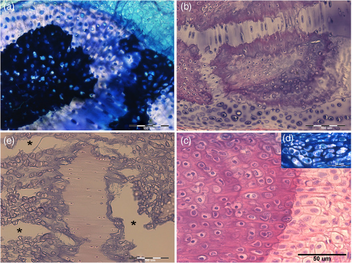
FIGURE 7.

Raja clavata (Chondrichthyes). Undecalcified, resin‐embedded sections of pelvic basipterygium radials stained with methylene‐blue/acid fuchsine. (a) (200x) Transverse section of cartilage calcifying zone showing a mix composition of densely mineralized and impending calcification zones. An uncalcified matrix is evident in the top right corner. (b) (200x) Stripes of uncalcified cartilage separating calcified zones. The chondrocytes are increasing volume, and the cell duplications develop an irregular distribution of tensional stresses in this mixed tissue, as documented by the morphology of the stripe above (stretched in the plane of the section) and that of the stripe below (where tensional forces have a different direction). (c) (100x) Fissures (asterisks) produced by tensional stresses in the recent cartilage calcified matrix and associated with stretching the uncalcified cartilage. In the first, chondrocyte lacunae show a trend to form rows along the strain force direction. (d) (400x) Detail of the calcification border showing the large chondrocyte lacunae (with recent cell duplications in the top right insert) and chondrocytes hypertrophy in the layer of uncalcified cartilage on the right.
