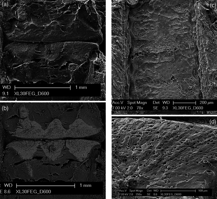
FIGURE 8.

Raja clavata (Chondrichthyes) scanning electron microscopy images of wing‐fin radials. (a–b) (secondary electron imaging (SEI) and BSE mode of the inter‐radial joint zone) The BSE image shows the mineralized joint disks with a tesseral‐like layout, forming a compact and flat disk on both sides. The calcified cartilage has a higher density of chondrocytes lacunae than the neighboring, uncalcified cartilage. (c) (SEI of mono‐columnar radial longitudinally sectioned above the calcified central column). Regular, low‐density distribution of chondrocyte lacunae. On both the right and left sides are evident several perichondrial layers. (d) (SEI of a multi‐columnar basiptergium transversally sectioned). Zone of early calcification with a high density of chondrocyte lacunae aligned in rows.
