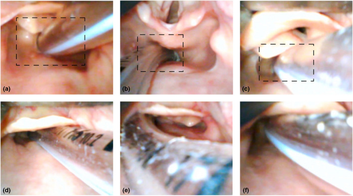
Figure 5.

Repeat laryngoscopy in the presence of oesophageal intubation. All tubes pictured are placed in the oesophagus. None meet the visual criteria for excluding oesophageal intubation on repeat laryngoscopy. The boxes (upper row) illustrate how a more restricted view, as might occur with challenging anatomy or use of direct laryngoscopy, could contribute to misinterpretation of the site of tube placement. This is particularly true if the practitioner is time pressured or at risk of confirmation bias. The arytenoids may be mistaken for the epiglottis (a and c), blanching of the lateral aspects of the oesophageal opening (c and f) or the cuff (b) may be mistaken for the vocal cords. The epiglottis may conceal the larynx entirely (D and F). In (e), the right arytenoid is visible lateral to the tube but cannot be confirmed to be passing posterior to it. All photos are clinical cadaver images from Dalhousie University's Human Body Donation Program. Used with permission.
