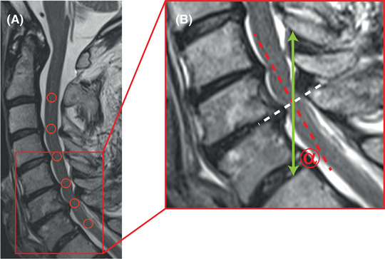
FIGURE 2.

Misalignment of the spinal cord in sagittal phase contrast MRI. In a patient with a multisegmental cervical spinal stenosis, the spinal cord shows a lordotic flection (A; midsagittal T2 weighted [w]; red circles representing phase contrast MRI [PC‐MRI] regions of interest). In axial imaging, the slice orientation was adjusted perpendicular to the spinal cord (B; midsagittal T2w, magnification of the red section in panel A, white dotted line representing the orientation of the axial PC‐MRI slice), measuring appropriately through plane craniocaudal spinal cord motion. In contrast, in sagittal PC‐MRI spinal cord motion was assessed craniocaudal within the field of view (B; midsagittal T2w; magnification of the red section in panel A; green arrow represents the orientation of the craniocaudal PC‐MRI velocity measurements), while the spinal cord was misaligned to the vertical reference line (B; sagittal T2w; magnification of the red section in panel A; dotted red line representing the spinal cord orientation at the level of the cervical disc C6/C7). The misalignment (B; sagittal T2w; magnification of the red section in panel A; misalignment angle α) may confound craniocaudal spinal cord motion measurements by a systematic measurement error.
