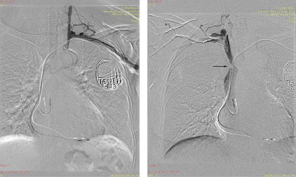
FIGURE 1.

Contrast venography of the left subclavian vein (left) and the right subclavian vein (right). Note the complete obstruction of the left innominate vein in the proximity of the PM leads. Left to right collateral is present. The arrow highlights the SVC stenosis [Colour figure can be viewed at wileyonlinelibrary.com]
