FIGURE 3.

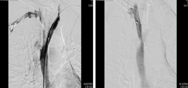
Contrasts venography after stent implantation showing a patent SVC and left innominate vein (left) and right innominate vein (right)

Contrasts venography after stent implantation showing a patent SVC and left innominate vein (left) and right innominate vein (right)