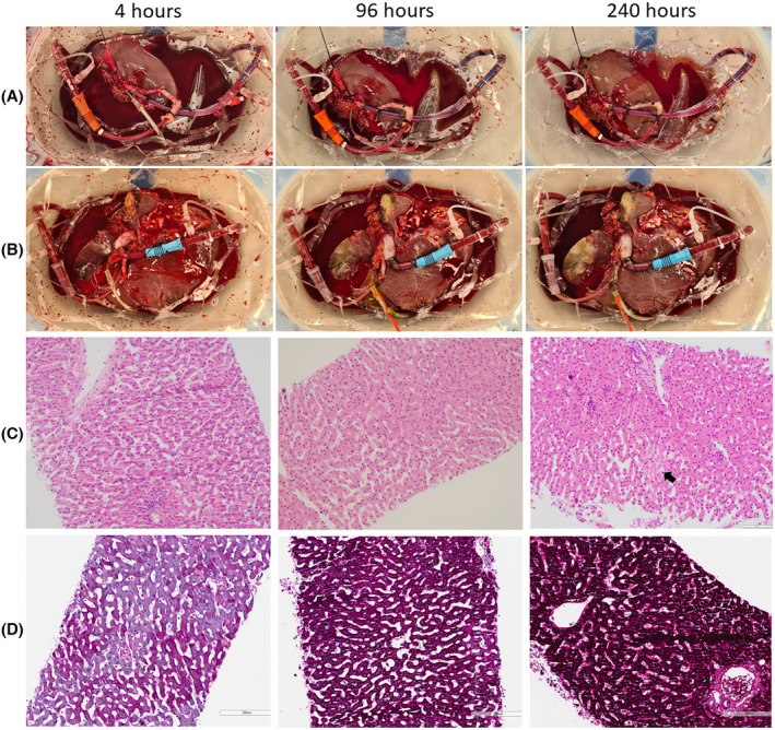
FIGURE 4.

Macroscopic and microscopic evidence of preserved architectural integrity during long‐term ex situ machine perfusion of human livers. Macroscopic images of left lateral segment grafts (A), and extended right grafts (B) at 4, 96, and 240 h after splitting. Hematoxylin and eosin‐stained sections of core biopsies (C) were taken at 4, 96, and 240 h after splitting (200× magnification). Occasional patchy areas of necrosis are seen (arrow) but the liver architecture was preserved in all sections from 4 h to 240 h after splitting (scale bar 100 μm). Periodic‐acid Schiff‐stained sections of core biopsies (D) were taken at 4, 96, and 240 h after splitting (200× magnification, scale bar 200 μm). Glycogen deposition increases within hepatocytes over time demonstrating energy storage during perfusion. [Color figure can be viewed at wileyonlinelibrary.com]
