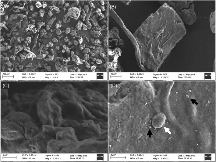
FIGURE 1.

SEM images of wMPs. wMPs morphology was analysed by SEM at low (A, B) and high magnification (C, D). Small dimension wMPs are visible on the surface of the bigger ones, including particles with a diameter of ≤2 μm (white arrow) and in the nanosized range (black arrows). Scale bars: (A) 100 μm; (B) 10 μm; (C and D) 2 μm
